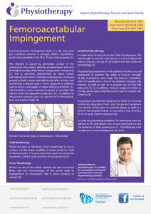
Femoracetabular Impingement

Femoroacetabular Impingement (FAI) is a hip and groin pain condition common in younger, athletic populations, particularly prevalent in the 18 to 35 year old age group [1].

This disorder is caused by premature contact of the acetabulum (hip socket) with the proximal head and neck (the ball of our hip) of the femur during certain movements [2]. FAI is generally characterised by deep seated, intermittent discomfort in the hip and groin areas, which can present as both a deep ache during and after activity, and occasionally a sharper pain in more aggravating positions such as sitting cross legged or when changing direction [3]. The condition is more common in sports or activities that require quick and repeated acceleration [4]. In addition to this primary symptom, pain can also be felt in the buttock, back and anterior thigh [3].

FAI has three main types of presentation, these being:

CAM Morphology

Where the neck of the femur (thigh bone) loses its round contour and thus loses its ability to rotate smoothly inside of the hip socket. This extra bone then abuts the socket on movement. CAM is move common in male patients [5].

Pincer Morphology

Where the rim of the socket extends out over its normal limits, and this ‘overcoverage’ of the socket leads to impingement on movement. This is more common in women [6].

Combined Morphology

A combination of both pincer and CAM morphologies. This was thought to be quite common but a recent longitudinal cohort study found only 2% of subjects had the combined morphology [5].

Physiotherapy assessment of FAI generally revolves around assessment of patients’ hip range of motion, strength of hip musculature and single leg balance. Individuals with FAI usually have a loss of hip abductor strength, hip rotators, as well as pain on a squeeze test of their hip adductors [1, 7]. In addition, reduced ranges of motion in the hip, particularly in hip flexion and internal rotation are common [8].

According to the Warwick Statement of 2016, for FAI to be confidently diagnosed there must be present symptoms and positive clinical signs as outlined above, as well as a positive radiological finding, so your physio may send you for an X-ray to confirm diagnosis [3].

As with any presenting condition, FAI treatment must be tailored to the individual’s own unique characteristics and the demands of their chosen pursuit. Physiotherapy aims to help you continue doing what you love!


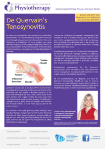
De Quervain’s Disease

What is De Quervain’s disease? This is a type of tenosynovitis in the wrist which is very common.

De Quervain’s tenosynovitis is an overload injury of tendons in the thumb. The two tendons that straighten the thumb travel through a tunnel (extensor retinaculum) in the wrist. Thickening or breakdown of these tendons can cause difficulty and pain when gliding through the tunnel. It is caused by repetitive and continuous strain of these tendons under the extensor retinaculum at the back (dorsal side) of the wrist.

Symptoms include pain at the base of the thumb which is exacerbated by movements of the wrist and thumb. Other symptoms can also include a burning sensation in the hand and swelling of the wrist. Pain is often made worse with activities that require turning the wrist away from the thumb (ulnar deviation) and gripping. De Quervain’s typically occurs in 30-50 years old females, new mothers, and manual workers. It is also more typically seen in the winter months.

De Quervain’s will begin gradually when starting a new activity or having a change in your normal routine. Provoking activities will slowly worsen symptoms and eventually you will get pain with most activities involving the hands. It is quite common for De Quervain’s tenosynovitis to affect both hands simultaneously.

One test for confirming De Quervain’s tenosynovitis is the Finkelstein’s test. It involves bending your thumb across your hand in a gripped position and turning the wrist away from the thumb. Pain along the base of the thumb, into the wrist and forearm is a positive result and is a strong indicator of De Quervain’s.

Physiotherapy treatment for De Quervain’s tenosynovitis can include ultrasound, massage, exercise, or splinting. Treatment will initially revolve around pain reduction and management. Once pain or other symptoms have resolved you will be prescribed exercises to gradually strengthen your thumbs to withstand load.

Physiotherapy management for De Quervain’s tenosynovitis is often successful in most cases. Sometimes further management including steroid injections or even surgery may be indicated but is quite rare.

All our physiotherapists can diagnose and treat De Quervain’s tenosynovitis. Should you have an issue in this area and need assistance, please don’t hesitate to contact either Berwick, Pakenham Lakeside or Clyde North Physiotherapy clinics to book in your next appointment.

Single Knee Bends

In this short video Chris explains how a simple movement such as standing single knee bend can tell a lot about someone’s biomechanics and potential issues that might be contributing or causing an injury. Chris is a little shy so no amount of asking nicely would get him on camera.

Adductor Strengthening Exercises – with Sejin An

Adductor strains are common on the sports field and in the work place. Often it can be due to an imbalance or weakness of the adductor muscles on the inside of the leg. In this short video, Se-Jin shows some easy progressive exercises to build strength in the adductor muscles.

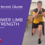
Lower Limb Strength – with Nathaniel Martin

In this video, Nathaniel shows us some variations to improve lower limb strength and build some functional strength for the pre season to help with running. It is very important to work on different positions of strength to improve control and function to maximise performance on the sports field and reduce the risk of injury.