Carpal Tunnel Syndrome

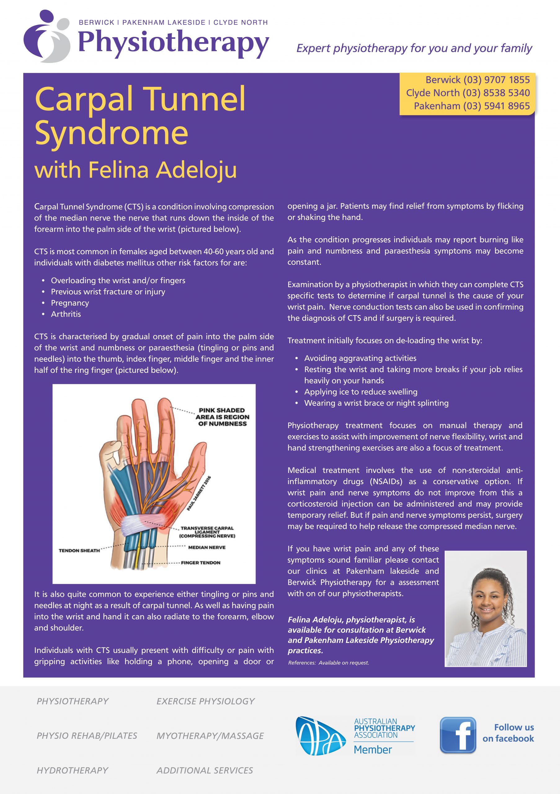
Carpal Tunnel Syndrome (CTS) is a condition involving compression of the median nerve the nerve that runs down the inside of the forearm into the palm side of the wrist (pictured below).

CTS is most common in females aged between 40-60 years old and individuals with diabetes mellitus other risk factors for are:

  • Overloading the wrist and/or fingers
  • Previous wrist fracture or injury
  • Pregnancy
  • Arthritis

CTS is characterised by gradual onset of pain into the palm side of the wrist and numbness or paraesthesia (tingling or pins and needles) into the thumb, index finger, middle finger and the inner half of the ring finger (pictured below).

It is also quite common to experience either tingling or pins and needles at night as a result of carpal tunnel. As well as having pain into the wrist and hand it can also radiate to the forearm, elbow and shoulder.

Individuals with CTS usually present with difficulty or pain with gripping activities like holding a phone, opening a door or opening a jar. Patients may find relief from symptoms by flicking or shaking the hand.

As the condition progresses individuals may report burning like pain and numbness and paraesthesia symptoms may become constant.

Examination by a physiotherapist in which they can complete CTS specific tests to determine if carpal tunnel is the cause of your wrist pain. Nerve conduction tests can also be used in confirming the diagnosis of CTS and if surgery is required.

Treatment initially focuses on de-loading the wrist by:

  • Avoiding aggravating activities
  • Resting the wrist and taking more breaks if your job relies heavily on your hands
  • Applying ice to reduce swelling
  • Wearing a wrist brace or night splinting

Physiotherapy treatment focuses on manual therapy and exercises to assist with improvement of nerve flexibility, wrist and hand strengthening exercises are also a focus of treatment.

Medical treatment involves the use of non-steroidal anti-inflammatory drugs (NSAIDs) as a conservative option. If wrist pain and nerve symptoms do not improve from this a corticosteroid injection can be administered and may provide temporary relief. But if pain and nerve symptoms persist, surgery may be required to help release the compressed median nerve.

If you have wrist pain and any of these symptoms sound familiar please contact our clinics at Pakenham lakeside and Berwick Physiotherapy for a assessment with on of our physiotherapists.

References:  Brukner, P. (2017). Brukner & Khan’s clinical sports medicine. (5th Edition) North Ryde: McGraw-Hill.  Genova, A., Dix, O., Saefan, A., Thakur, M., & Hassan, A. (2020). Carpal tunnel syndrome: a review of literature. Cureus12(3).  Chesterton, L. S., Blagojevic-Bucknall, M., Burton, C., Dziedzic, K. S., Davenport, G., Jowett, S. M., … & Roddy, E. (2018). The clinical and cost-effectiveness of corticosteroid injection versus night splints for carpal tunnel syndrome (INSTINCTS trial): an open-label, parallel group, randomised controlled trial. The Lancet392(10156), 1423-1433.  Bobowik, P. Ż. (2019). Effectiveness of physiotherapy in carpal tunnel syndrome (CTS). Postępy Rehabilitacji, 2019(2), 47-58. doi:10.5114/areh.2019.8502


Frozen Shoulder

Have you noticed of late a gradual increase of stiffness and/or pain in your shoulder, sometimes without any obvious mechanism of injury? Wondering why on earth lying on one side of your body at night is agony, and basic tasks like getting dressed or hanging the washing on the line are getting harder and harder?

Frozen Shoulder, referred to as Adhesive Capsulitis, is a condition characterised by painful and/or restricted shoulder movement [1]. The condition involves the shoulder capsule (strong band of ligaments that surround our shoulder joint) becoming thickened and suffering contracture. There is currently some debate over whether the condition is due to inflammation, fibrosis of the capsule, or both [2].

Whilst most cases of Frozen Shoulder are idiopathic (no known cause), cases of Frozen Shoulder can be seen more commonly in people with type 2 diabetes, thyroid disorders, those who have had recently been immobilised after shoulder surgery or have suffered a recent traumatic fall, recent stroke and cancer sufferers, as well as a previous history of Dupuytren’s contracture [3, 4].

The condition is far more common in those over the age of 40, with the most common age group being 40-60 year olds. In addition, Women are more likely than Men to suffer the condition [5].

There are three main stages of Frozen Shoulder, being:

  • Freezing – The ‘Freezing’ stage or ‘Pain over Stiffness’ is usually characterised by sharp pain with movement in the shoulder joint, with progressively worsening movement.
  • Frozen – This phase, also known as the ‘Stiffness over Pain’ phase where pain may diminish, however stiffness usually plateaus or could get slightly worse.
  • Thawing – This final phase involves the progressive increase of movement and reduction of pain [6].

In all the condition generally lasts for between 9-18 months, but can last from 5-24 months [6]. Diagnosis is usually formed by a mixture of symptoms, physical range and strength assessment and imaging [7,8].

Treatment for Frozen Shoulder in our clinic can vary but can involve gentle range, stretching and strengthening exercises, complimented by joint mobilisations and soft tissue release.

Common exercises for adhesive capsulitis include pendular movements, crawling hands up the wall, doorframe and child’s pose stretch, and the use of shoulder pulleys, to gently encourage further range in the affected shoulder by using the non-affected shoulder.

In addition to this, we have seen some success in pain and movement improvements with a short course of prednisolone (oral cortisone). In more severe cases, treatment options can include both corticosteroid injections into the shoulder, or a hydro-dilatation, which is where a combination of saline and cortisone is injected into the capsule in order to distend it, and ‘break’ the contractures in the capsule [9]. Research has shown that initial conservative treatments such as these can be effective in 90% of patients, without the need for surgical intervention [9].

Whilst Frozen Shoulder is at times a scary and frustrating condition, with effective and efficient physiotherapy your symptoms can be managed well, and our physios are extremely well placed to guide you through this. If you think the above condition sounds like you, be sure to come down and see one of our expert therapists for an assessment ASAP!

References:  [1] Dias R, Cutts S, Massoud S. Frozen shoulder. Bmj. 2005 Dec 15;331(7530):1453-6;  [2] Cho CH, Song KS, Kim BS, Kim DH, Lho YM. Biological aspect of pathophysiology for frozen shoulder. BioMed research international. 2018 May 24;2018; [3] Mezian K, Chang KV. Frozen Shoulder. StatPearls [Internet]. 2019 Feb 25; [4] Whelton C, Peach CA. Review of diabetic frozen shoulder. European Journal of Orthopaedic Surgery & Traumatology. 2018 Apr;28(3):363-71; [5] Uppal HS, Evans JP, Smith C. Frozen shoulder: a systematic review of therapeutic options. World journal of orthopedics. 2015 Mar 18;6(2):263; [6] Chan HB, Pua PY, How CH. Physical therapy in the management of frozen shoulder. Singapore medical journal. 2017 Dec;58(12):685;  [7] Kelley MJ, Shaffer MA, Kuhn JE, Michener LA, Seitz AL, Uhl TL, Godges JJ, McClure PW, Altman RD, Davenport T, Davies GJ. Shoulder pain and mobility deficits: adhesive capsulitis: clinical practice guidelines linked to the international classification of functioning, disability, and health from the Orthopaedic Section of the American Physical Therapy Association. Journal of orthopaedic & sports physical therapy. 2013 May;43(5):A1-31;  [8] Li JQ, Tang KL, Wang J, Li QY, Xu HT, Yang HF, Tan LW, Liu KJ, Zhang SX. MRI findings for frozen shoulder evaluation: is the thickness of the coracohumeral ligament a valuable diagnostic tool?. PLoS One. 2011 Dec 7;6(12):e28704;  [9] Cho CH, Bae KC, Kim DH. Treatment strategy for frozen shoulder. Clinics in orthopedic surgery. 2019 Sep;11(3):249.


Femoracetabular Impingement

Femoroacetabular Impingement (FAI) is a hip and groin pain condition common in younger, athletic populations, particularly prevalent in the 18 to 35 year old age group [1].

This disorder is caused by premature contact of the acetabulum (hip socket) with the proximal head and neck (the ball of our hip) of the femur during certain movements [2]. FAI is generally characterised by deep seated, intermittent discomfort in the hip and groin areas, which can present as both a deep ache during and after activity, and occasionally a sharper pain in more aggravating positions such as sitting cross legged or when changing direction [3]. The condition is more common in sports or activities that require quick and repeated acceleration [4]. In addition to this primary symptom, pain can also be felt in the buttock, back and anterior thigh [3].

FAI has three main types of presentation, these being:

CAM Morphology

Where the neck of the femur (thigh bone) loses its round contour and thus loses its ability to rotate smoothly inside of the hip socket. This extra bone then abuts the socket on movement. CAM is move common in male patients [5].

Pincer Morphology

Where the rim of the socket extends out over its normal limits, and this ‘overcoverage’ of the socket leads to impingement on movement. This is more common in women [6].

Combined Morphology

A combination of both pincer and CAM morphologies. This was thought to be quite common but a recent longitudinal cohort study found only 2% of subjects had the combined morphology [5].

Physiotherapy assessment of FAI generally revolves around assessment of patients’ hip range of motion, strength of hip musculature and single leg balance. Individuals with FAI usually have a loss of hip abductor strength, hip rotators, as well as pain on a squeeze test of their hip adductors [1, 7]. In addition, reduced ranges of motion in the hip, particularly in hip flexion and internal rotation are common [8].

According to the Warwick Statement of 2016, for FAI to be confidently diagnosed there must be present symptoms and positive clinical signs as outlined above, as well as a positive radiological finding, so your physio may send you for an X-ray to confirm diagnosis [3].

As with any presenting condition, FAI treatment must be tailored to the individual’s own unique characteristics and the demands of their chosen pursuit. Physiotherapy aims to help you continue doing what you love!

De Quervain’s Disease

What is De Quervain’s disease? This is a type of tenosynovitis in the wrist which is very common.

De Quervain’s tenosynovitis is an overload injury of tendons in the thumb. The two tendons that straighten the thumb travel through a tunnel (extensor retinaculum) in the wrist. Thickening or breakdown of these tendons can cause difficulty and pain when gliding through the tunnel. It is caused by repetitive and continuous strain of these tendons under the extensor retinaculum at the back (dorsal side) of the wrist.

Symptoms include pain at the base of the thumb which is exacerbated by movements of the wrist and thumb. Other symptoms can also include a burning sensation in the hand and swelling of the wrist. Pain is often made worse with activities that require turning the wrist away from the thumb (ulnar deviation) and gripping. De Quervain’s typically occurs in 30-50 years old females, new mothers, and manual workers. It is also more typically seen in the winter months.

De Quervain’s will begin gradually when starting a new activity or having a change in your normal routine. Provoking activities will slowly worsen symptoms and eventually you will get pain with most activities involving the hands. It is quite common for De Quervain’s tenosynovitis to affect both hands simultaneously.

One test for confirming De Quervain’s tenosynovitis is the Finkelstein’s test. It involves bending your thumb across your hand in a gripped position and turning the wrist away from the thumb. Pain along the base of the thumb, into the wrist and forearm is a positive result and is a strong indicator of De Quervain’s.

Physiotherapy treatment for De Quervain’s tenosynovitis can include ultrasound, massage, exercise, or splinting. Treatment will initially revolve around pain reduction and management. Once pain or other symptoms have resolved you will be prescribed exercises to gradually strengthen your thumbs to withstand load.

Physiotherapy management for De Quervain’s tenosynovitis is often successful in most cases. Sometimes further management including steroid injections or even surgery may be indicated but is quite rare.

All our physiotherapists can diagnose and treat De Quervain’s tenosynovitis. Should you have an issue in this area and need assistance, please don’t hesitate to contact either Berwick, Pakenham Lakeside or Clyde North Physiotherapy clinics to book in your next appointment.

Tennis Elbow

Tennis elbow (also known as lateral epicondylitis) is a common overuse injury causing pain into the outer (lateral) side of the elbow.

Although the name suggests the cause of this pain to be as a result of playing tennis, less than 5% of cases are as a result of this. More common causes include repetitive activities like computer use, heavy lifting and activities requiring repeated twisting at the wrist (common for electricians, carpenters and gardeners). These repetitive activities result in an excessive amount of strain through the tendons and muscles of the outer forearm. This overuse causes degenerative changes to occur at a cellular level in the tendon leading to a reduction in the tendon’s ability to tolerate load.

Common symptoms are tenderness over the bony prominence of the lateral elbow (lateral epicondyle), and pain that radiates up the arm or down the outside of the forearm. Individuals may also experience pain and reduced strength with activities such as lifting or carrying in the affected arm, and with gripping activities, for example, opening jars and opening doors. Pain may also be provoked with activities involving twisting at the wrist or positions where the palm is turned face down, such as using a screwdriver or typing at a computer.

A common indicator of tennis elbow is that, initially, pain will begin after the provoking activity, and then slowly progress to pain during the aggravating activity, to then having pain constantly (in the later stages of the injury) which will limit you from being able to complete other activities beside the initially aggravating one.

Physiotherapy treatment usually involves an initial focus on reducing pain and irritation with a combination of manual therapy techniques such as soft tissue massage, ultrasound and exercises to gently stretch the muscles of the forearm.

The focus of treatment in the management of tennis elbow is to reduce the excessive loading through the tendon and progressively rebuild the tendon’s tolerance to load. This is achieved through a graduated loading program which your physiotherapist will prescribe for you.

Use of a tennis elbow brace can sometimes be extremely effective in managing the load through the tendons of the lateral elbow, especially in individuals who are unable or have difficulty reducing aggravating activities. Our physiotherapists are experienced in fitting these and will advise you as to whether a brace would be beneficial, and if so, how and when to wear it.

Physiotherapy management for tennis elbow is successful in most cases. Certain cases that do not respond to initial treatment may require further investigation, and possibly medical intervention if it is indicated.

All our physiotherapists are skilled in the diagnosis and management of tennis elbow. Should you need some help in this area, don’t hesitate to contact either our Berwick or Pakenham Lakeside Physiotherapy clinic to book in your appointment.

Achilles Tendinopathy

ACHILLES PAIN STOPPING YOU EXERCISING? Started really increasing your running load during this time of COVID-19 shutdown, but have been plagued by an unusual pain in the back of your lower leg? You are not alone!…Click here to find out more…

As the world adjusts to this world of closed gyms and reduced access to equipment, many people are lacing up their runners and hitting the pavement. This is a great thing! I love running, and have a special interest in the management of runners’ injuries.

As this has increased, however, we have seen an increase of people in the clinic with classic runners-based injuries, such as anterior knee pain, and Achilles tendiopathy.

Your Achilles Tendon is the large, strong band of tissue that attaches the bottom of your calf muscle to your calcaneus (heel bone). It is critical in running, as it helps absorb force as we land, and then helps propel us forward as we go up onto our toes to move onto our next stride.

Achilles Tendinopathy, previously (and still commonly) known as Achilles Tendinitis, is a condition where the tendon suffers a failed healing response to load, and the collagen fibres which make up the majority of the tendon are disrupted [1]. This change in the makeup of the tendon can leave it more susceptible to pain and damage. It is not, as previously thought, a purely inflammatory condition.

The question is how to prevent it, and then treat it once it’s here? As someone who suffered from AT throughout my footballing career and had it occasionally pop back up since I started running, I can say the ability to correctly manage your load (the distance, intensity and time you run for) is paramount in correctly dealing with AT. Our physios are experts at developing and personalising loading programs, and can help you through this process.

Secondly, it is absolutely vital that anyone who is running has a resistance training program that compliments it. One of the primary clinical risk factors of developing AT is a lack of strength in our calves, which makes sense as it’s the muscle that the Achilles originates from! [2] This doesn’t mean pounding massive weights and complicated exercises! Effective resistance exercises for runners can be done easily with body weight or extremely minimal equipment, and we are expertly placed to help you with this. A structured strengthening program that progressively loads the tendon is an effective treatment strategy at both reducing pain and increasing function in individuals with AT [3].

Finally, as with any running injury, ensuring that you have correct footwear that is appropriate for your individual needs is critical. Poor foot mechanics can put forces on the tendon that it cannot handle easily, which can increase the likelihood of developing the condition.

So, if you plan on hitting the pavement again and unsure about how to go about it, or are experiencing some Achilles pain, come on down to our Berwick or Pakenham Lakeside clinics, and let us keep you enjoying your running pain-free!

Suffering from leg pain when running?

Working Remotely from Home

COVID-19 NECK & BACK – 2020’s newest musculoskeletal condition!

Have you been working from home and are now starting to experience some neck and back pain? The good news is there are strategies to help manage and reduce your pain!

The success of a remote working arrangement is often dependent upon a number of factors. Whilst the office is often designed to aid productivity and provide a safe workstation, many homes lack these features. This document is to assist you in optimising your remote working set up and to reduce your risk of musculoskeletal pain or injury. Regular exercise should remain or become an essential element of your day. This does not mean running or lifting heavy weights, but could take the form of a brisk walk and some gentle stretching.

Movement is Medicine!

Sit less, move more:

Recent research has shown long periods of sitting (regardless of exercise regime) can lead to increased risk of preventable musculoskeletal disorders and chronic diseases such as diabetes. Make sure you get out of your chair and leave your workstation every 20-30 minutes. If you find that you lose track of time while working, set a countdown timer on your phone to assist.

Reduce eye strain:

To control visual fatigue, a change of focus, such as a view out of a window or to a picture along a hallway, can provide exercise to focal muscles of the eyes whilst resting the tired muscles. Resting your eyes is not flicking on Facebook or Instagram!

Break up Repetitive tasks:

Timetable your day to allow for scheduled breaks and changes to repetitive tasks

Download our infographics for further information.

Working Remotely or From Home

Single Knee Bends

In this short video Chris explains how a simple movement such as standing single knee bend can tell a lot about someone’s biomechanics and potential issues that might be contributing or causing an injury. Chris is a little shy so no amount of asking nicely would get him on camera.

Adductor Strengthening Exercises – with Sejin An

Adductor strains are common on the sports field and in the work place. Often it can be due to an imbalance or weakness of the adductor muscles on the inside of the leg. In this short video, Se-Jin shows some easy progressive exercises to build strength in the adductor muscles.

Lower Limb Strength – with Nathaniel Martin

In this video, Nathaniel shows us some variations to improve lower limb strength and build some functional strength for the pre season to help with running. It is very important to work on different positions of strength to improve control and function to maximise performance on the sports field and reduce the risk of injury.