Shin Splints

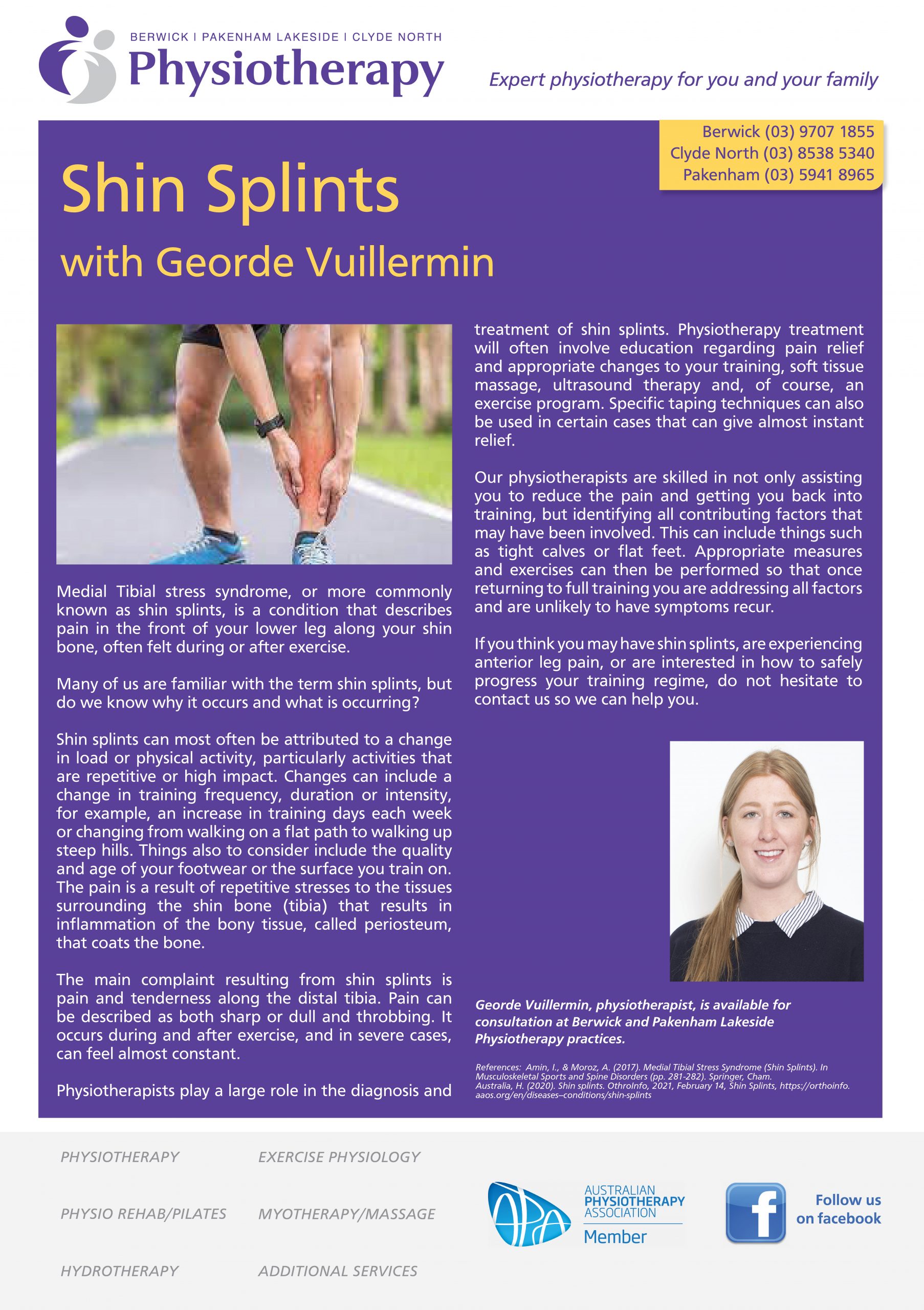
Medial Tibial stress syndrome, or more commonly known as shin splints, is a condition that describes pain in the front of your lower leg along your shin bone, often felt during or after exercise.

Many of us are familiar with the term shin splints, but do we know why it occurs and what is occurring?

Shin splints can most often be attributed to a change in load or physical activity, particularly activities that are repetitive or high impact. Changes can include a change in training frequency, duration or intensity. For example, an increase in training days each week or changing from walking on a flat path to walking up steep hills. Things also to consider include the quality and age of your footwear or the surface you train on.

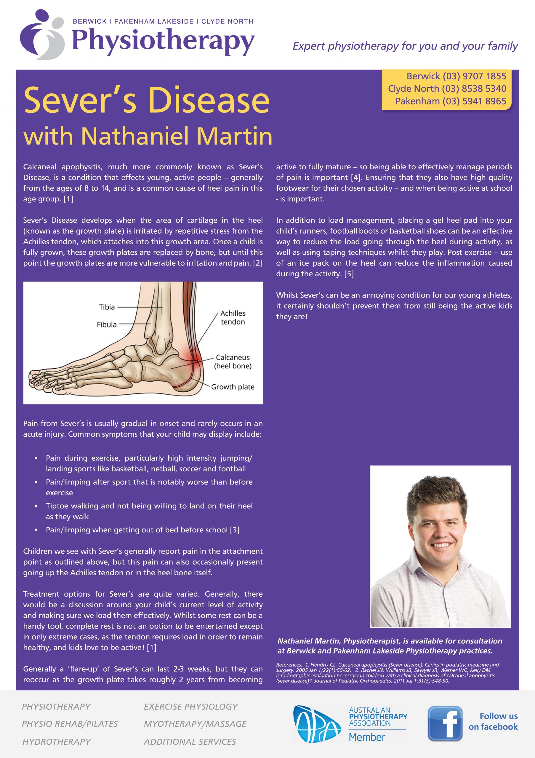
The pain is a result of repetitive stresses to the tissues surrounding the shin bone (tibia) that results in inflammation of the bony tissue, called periosteum, that coats the bone.

The main complaint resulting from shin splints is pain and tenderness along the distal tibia. Pain can be described as both sharp or dull and throbbing. It occurs during and after exercise and in severe cases can feel almost constant.

Physiotherapists play a large role in the diagnosis and treatment of shin splints. Physiotherapy treatment will often involve education regarding pain relief and appropriate changes to your training, soft tissue massage, ultrasound therapy, and of course an exercise program. Specific taping techniques can also be used in certain cases that can give almost instant relief.

Our Physios are skilled in not only assisting you to reduce the pain and getting you back into training, but identifying all contributing factors that may have been involved. This can include things such as tight calves or flat feet. Appropriate measures and exercises can then be performed so that once returning to full training you are addressing all factors and unlikely to have symptoms recur.

If you think you may have shin splints, are experiencing anterior leg pain or are interested in how to safely progress your training regime, do not hesitate to contact us so we can help you.

References:
Amin, I., & Moroz, A. (2017). Medial Tibial Stress Syndrome (Shin Splints). In Musculoskeletal Sports and Spine Disorders (pp. 281-282). Springer, Cham.  Australia, H. (2020). Shin splints.  OthroInfo, 2021, February 14, Shin Splints, https://orthoinfo.aaos.org/en/diseases–conditions/shin-splints


Hamstring Strains and Prevention

As many of us return sport this year after a year-long hiatus thanks to COVID-19, injury prevention should be something at the forefront of our minds – in particular, hamstring injury prevention.

Hamstrings strains are one of the most common sporting injuries. They occur frequently in sports that involve high speed movement including football, field hockey, cricket and running, to name a few. Hamstring strains have one of the highest re-injury numbers especially in soccer, football and rugby.

There are two types of hamstring strains: Type 1 hamstrings injuries commonly occur during fast pace running e.g sprinting or in football and commonly involve the bicep femoris muscle which is seen in the image below; Type 2 injuries are due to over stretching activities usually with the knee fully extended for example high kicking or during the splits. These injuries may occur at slow speed and commonly involve the semimembranosus muscle, also pictured below.

Hamstring strains commonly present with sudden onset of pain into the hamstring region. This pain usually prevents continuation of the activity that has caused the injury. In a type 1 hamstring injury, pain is generally located in the bicep femoris muscle, whereas in a type 2 hamstring injury, pain is usually located near the ischial tuberosity (sit bone) where the hamstring inserts into the bone.
On examination, they are tender over the location of the tear and may present with reduced strength on knee flexion (bending the knee) and have pain on knee extension (straightening the knee out) due to stretching the damaged muscle fibres. In high grade tears, bruising and inflammation can somtimes present in the posterior thigh.

Imaging through ultrasound and MRI can also be used to determine the severity and location of the hamstring strain if it is required by your physiotherapist.

Early management includes a rest, ice, compression and elevation program (RICE principle). After the initial management, the focus is to normalise walking and regain the strength and flexibility in the hamstring. This is achieved by implementing a graduated hamstring strengthening program.

Education on the recurrence of hamstring injuries is important as you are seven times more likely to re-injure a hamstring after a strain. Your physiotherapist can provide you with a hamstring injury prevention program if you play sport and it is highly recommended in athletes that participate in kicking and running sports.

The general recovery time frame for a hamstring strain is between 4-8 weeks, however this is dependent on the initial severity of the injury and the individual’s rate of recovery.

If you have hamstring pain or have had a hamstring strain in the past and want more information on hamstring rehab and specific exercises for a hamstring injury prevention program either for yourself, sporting group or club, please get in contact with one of highly qualified physiotherapists. They are all extremely competent in hamstring strain diagnosis and management, and are available for consultation at our Berwick, Clyde North and Pakenham Lakeside Physiotherapy clinics.


References:  Brukner, P. (2017). Brukner & Khan’s clinical sports medicine. (5th Edition) North Ryde: McGraw-Hill.  Ekstrand, J., Hägglund, M., & Waldén, M. (2011). Epidemiology of muscle injuries in professional football (soccer). The American journal of sports medicine, 39(6), 1226-1232.  Erickson, L. N., & Sherry, M. A. (2017). Rehabilitation and return to sport after hamstring strain injury. Journal of Sport and Health Science

Vertigo – Dizziness and the Vestibular System with Ross de Valle

Dizziness can be an extremely debilitating symptom and unfortunately is surprisingly common. It is estimated that 15% of the population will experience dizziness at some stage of their life. One of the primary causes of dizziness is dysfunction of the vestibular system.

The vestibular system is part of the inner ear and brain which monitors the position and motion of the head. It is essential in maintaining visual stability, postural stability, and perception of movement. The vestibular apparatus is part of the inner ear and is approximately the size of a 5-cent piece.

Disorders of the vestibular system affect an individual’s balance and often impact their ability to walk, run and even drive a car. Benign Paroxysmal Positional Vertigo (BPPV) is the most common vestibular disorder and is thought to account for up to 42% of all vestibular disorders (Bhattacharyya et al CPG 2017).

  • Benign – it is not life-threatening.
  • Paroxysmal – it comes in sudden, brief spells.
  • Positional – it gets triggered by certain head
    positions or movements.
  • Vertigo – a false sense of movement.

Luckily, physiotherapy can help in the treatment of vestibular disorders. A thorough assessment is vital to determine if the vestibular system is the cause of the presenting symptoms. This also allows appropriate treatment which may include ‘canal repositioning manoeuvres’ which are used to treat BPPV or a targeted rehabilitation program for cases of vestibular dysfunction due to other causes.

Ross de Valle has a special interest in the treatment of vestibular disorders. He has completed additional training in vestibular rehabilitation and is available for consultation at both our Berwick and Clyde North clinics.

References:  Bhattacharyya N et al. Clinical Practice Guidelines: Benign Paroxysmal Positional Vertigo (Update) Otolaryngol Head Neck Surgery. 2017; 156 (3_suppl): S1-S47

Physiotherapy Management of Tendon Injuries

Tendon injuries are seen commonly by our Physiotherapy team. Often injuries to a tendon are related to an increase in load which results in changes to the structure of the tendon, causing pain and dysfunction. Injuries such as tennis elbow, Patellar tendinopathy and Achilles tendinopathy can occur when a patient starts a new type of activity or increases the amount of load to a current activity. There is a limit to the adaptive nature of tendons and this can result in a repetitive strain or overload tendon injury.

Evidence based research has shown that one form of management stands above all types of treatment of tendon injuries. This treatment involves a finely balanced understanding of tendon structure and biomechanics.

The key for managing tendon injury is understanding the threshold of the tendon and it’s ability to withstand load. When a patient injures a tendon, it is important to manage the amount of load on the tendon to reduce overload, but it is equally important to find a way to continue to load the tendon under it’s threshold, to allow it to heal and strengthen over time. Tendons want and need to be loaded in the right way. Load is how a tendon grows and heals. Without any load, they weaken and become more susceptible to ongoing trauma and injury which becomes a perpetual problem often seen in chronic tendon injury. This is the complexity of tendon injuries. Some rest is important, but not too much. Some load is important, but not too much. Our physiotherapists at First Choice Health are experts in understanding tendon pathology and implementing plans to allow adaptive healing and strengthening of tendons to enable recovery from repetitive injuries.

By using strategies to improve technique and biomechanics, and strength exercises such as isometric and eccentric loading, tendon injuries can be managed and improved over time. Expert advice and a thorough strength plan is required to overcome tendon injuries so that you can return to all activities pain free and stronger than ever before.

If you have a niggly tendon injury or some tendon pain that won’t go away, give our clinic a call to start your path to recovery.

Sever’s Disease

Calcaneal apophysitis, much more commonly known as Sever’s Disease, is a condition that effects young, active people – generally from the ages of 8 to 14, and is a common cause of heel pain in this age group. [1]

Sever’s Disease develops when the area of cartilage in the heel (known as the growth plate) is irritated by repetitive stress from the Achilles tendon, which attaches into this growth area. Once a child is fully grown, these growth plates are replaced by bone, but until this point the growth plates are more vulnerable to irritation and pain. [2]

Pain from Sever’s is usually gradual in onset and rarely occurs in an acute injury. Common symptoms that your child may display include:

  • Pain during exercise, particularly high intensity jumping/landing sports like basketball, netball, soccer and football
  • Pain/limping after sport that is notably worse than before exercise
  • Tiptoe walking and not being willing to land on their heel as they walk
  • Pain/limping when getting out of bed before school [3]

Children we see with Sever’s generally report pain in the attachment point as outlined above, but this pain can also occasionally present going up the Achilles tendon or in the heel bone itself.

Treatment options for Sever’s are quite varied. Generally, there would be a discussion around your child’s current level of activity and making sure we load them effectively. Whilst some rest can be a handy tool, complete rest is not an option to be entertained except in only extreme cases, as the tendon requires load in order to remain healthy, and kids love to be active! [1]

Generally a ‘flare-up’ of Sever’s can last 2-3 weeks, but they can reoccur as the growth plate takes roughly 2 years from becoming active to fully mature – so being able to effectively manage periods of pain is important [4]. Ensuring that they also have high quality footwear for their chosen activity – and when being active at school – is important.

In addition to load management, placing a gel heel pad into your child’s runners, football boots or basketball shoes can be an effective way to reduce the load going through the heel during activity, as well as using taping techniques whilst they play. Post exercise – use of an ice pack on the heel can reduce the inflammation caused during the activity. [5]

Whilst Sever’s can be an annoying condition for our young athletes, it certainly shouldn’t prevent them from still being the active kids they are!

References:  1. Hendrix CL. Calcaneal apophysitis (Sever disease). Clinics in podiatric medicine and surgery. 2005 Jan 1;22(1):55-62; 2. Rachel JN, Williams JB, Sawyer JR, Warner WC, Kelly DM. Is radiographic evaluation necessary in children with a clinical diagnosis of calcaneal apophysitis (sever disease)?. Journal of Pediatric Orthopaedics. 2011 Jul 1;31(5):548-50; 3. Bailey CW, Cannon ML. Sever disease (calcaneal apophysitis). Journal of Osteopathic Medicine. 2014 May 1;114(5):411-; 4. Madden CC, Mellion MB. Sever’s disease and other causes of heel pain in adolescents. American family physician. 1996 Nov 1;54(6):1995-2000; 5. Smith JM, Varacallo M. Sever disease. 2017

Meniscus Injuries

Meniscus injuries are one of the most common injuries of the knee. The meniscus refers to two wedge shaped pieces of fibrocartilage in your knee joint between your tibia and femur. They have an important role in distributing load and act as a shock absorber within the knee joint.

Meniscus injuries can be degenerative or occur as a result of an acute injury. Acute meniscus tears during sport often involve a quick twisting of the upper leg on a slightly flexed knee whilst the foot stays planted. It can be with or without direct contact. In sport they can often occur alongside other injuries such as an anterior cruciate ligament (ACL) injury, so it is important to see a health professional for an appropriate diagnosis.

In the case of a degenerative meniscus tear an awkward twist getting out of the car or stepping down a step may be enough to cause a tear.

When injuring your meniscus, common symptoms include pain localised to the side of the joint (either side depending if the medial or lateral meniscus is involved) when you try to bend, straighten or twist the knee. Swelling can occur and in some cases, you may here clicking, popping or feel as if the knee is locking, as the cartilage may be catching between the joint surfaces and blocking full range of movement.

Your Physiotherapist can assist in the initial assessment and diagnosis of a knee injury and in many cases if a meniscus injury is suspected you will be referred for imaging. Depending on the classification of the tear (size and location) and your presenting symptoms (pain, locking, clicking) surgical management may be necessary.

Physiotherapy is an integral part of the rehabilitation process after a meniscus injury regardless whether treatment is conservative or surgical. Different treatment techniques are used depending on the stage of your rehabilitation. Initially, treatment may involve manual therapy such as massage and education for pain relief, and progress gradually to functional activities including strengthening and neuromuscular retraining exercises to assist your safe return to your previous levels of activity.

If you suspect you have a meniscus injury make sure to contact us and we can help with the initial diagnosis and support you through the rehabilitation process.

References: OthroInfo, 2021, February 16, Meniscus Tears, https://orthoinfo.aaos.org/en/diseases–conditions/meniscus-tears/ Sports Medicine Australia, 2021, February 16, Meniscus Injury A guide to prevention and management, https://sma.org.au/sma-site-content/uploads/2013/05/719-SMA-InjuryBrochure-Meniscus_web.pdf

Plantar Fasciitis

Have you recently noticed a sharp, burning or stabbing pain into the bottom of your foot/heel? Are you noticing it is most uncomfortable when you first get out of bed and walk to the kitchen? If so, you could be suffering from a common inflammatory condition known as plantar fasciitis (pronounced plantar fas-ci-i-tis).

The main symptom associated with plantar fasciitis is pain experienced first thing in the morning, following prolonged periods of rest or sedentary positions and also after extended periods of exercise or standing. Pain is usually located in the heel and arch of the foot, but can occasionally be located into the ball of the foot as well.

The plantar fascia is the thick fibrous tissue on the underside of the foot, starting from the calcaneus (heel bone) and connects along the sole to the ball of your foot. It acts like a rubber band by stretching and contracting as you walk. The plantar fascia can be put under quite a bit of force, particularly when participating in activities with running or jumping movement.

Plantar fasciitis is diagnosed when this soft tissue is inflamed.  This can occur following an acute injury, however it most commonly develops due to overuse.

Factors of overuse plantar fasciitis include:

  • A sudden increase in activity/exercise load
  • Weight gain
  • Poor footwear
  • Poor biomechanics (excessive pronation)

Physiotherapy treatment usually involves initially reducing pain and inflammation. This is done through activity modification, footwear advice and using manual therapy such as soft tissue massage, ultrasound and strapping the arch of the foot.

Our physiotherapists can provide a graded exercise program focusing on stretching and strengthening the soft tissue of the foot and surrounding musculature. Our physiotherapists can also assist in prescribing arch supporting orthotics, if they believe this is indicated.

All of our physiotherapists are skilled in diagnosing and treating plantar fasciitis.  If you have been suffering with this condition and would like some help, don’t hesitate to contact any of our Berwick, Pakenham Lakeside or Clyde North physiotherapy clinics to book an appointment.

Femoracetabular Impingement

Femoroacetabular Impingement (FAI) is a hip and groin pain condition common in younger, athletic populations, particularly prevalent in the 18 to 35 year old age group [1].

This disorder is caused by premature contact of the acetabulum (hip socket) with the proximal head and neck (the ball of our hip) of the femur during certain movements [2]. FAI is generally characterised by deep seated, intermittent discomfort in the hip and groin areas, which can present as both a deep ache during and after activity, and occasionally a sharper pain in more aggravating positions such as sitting cross legged or when changing direction [3]. The condition is more common in sports or activities that require quick and repeated acceleration [4]. In addition to this primary symptom, pain can also be felt in the buttock, back and anterior thigh [3].

FAI has three main types of presentation, these being:

CAM Morphology

Where the neck of the femur (thigh bone) loses its round contour and thus loses its ability to rotate smoothly inside of the hip socket. This extra bone then abuts the socket on movement. CAM is move common in male patients [5].

Pincer Morphology

Where the rim of the socket extends out over its normal limits, and this ‘overcoverage’ of the socket leads to impingement on movement. This is more common in women [6].

Combined Morphology

A combination of both pincer and CAM morphologies. This was thought to be quite common but a recent longitudinal cohort study found only 2% of subjects had the combined morphology [5].

Physiotherapy assessment of FAI generally revolves around assessment of patients’ hip range of motion, strength of hip musculature and single leg balance. Individuals with FAI usually have a loss of hip abductor strength, hip rotators, as well as pain on a squeeze test of their hip adductors [1, 7]. In addition, reduced ranges of motion in the hip, particularly in hip flexion and internal rotation are common [8].

According to the Warwick Statement of 2016, for FAI to be confidently diagnosed there must be present symptoms and positive clinical signs as outlined above, as well as a positive radiological finding, so your physio may send you for an X-ray to confirm diagnosis [3].

As with any presenting condition, FAI treatment must be tailored to the individual’s own unique characteristics and the demands of their chosen pursuit. Physiotherapy aims to help you continue doing what you love!

De Quervain’s Disease

What is De Quervain’s disease? This is a type of tenosynovitis in the wrist which is very common.

De Quervain’s tenosynovitis is an overload injury of tendons in the thumb. The two tendons that straighten the thumb travel through a tunnel (extensor retinaculum) in the wrist. Thickening or breakdown of these tendons can cause difficulty and pain when gliding through the tunnel. It is caused by repetitive and continuous strain of these tendons under the extensor retinaculum at the back (dorsal side) of the wrist.

Symptoms include pain at the base of the thumb which is exacerbated by movements of the wrist and thumb. Other symptoms can also include a burning sensation in the hand and swelling of the wrist. Pain is often made worse with activities that require turning the wrist away from the thumb (ulnar deviation) and gripping. De Quervain’s typically occurs in 30-50 years old females, new mothers, and manual workers. It is also more typically seen in the winter months.

De Quervain’s will begin gradually when starting a new activity or having a change in your normal routine. Provoking activities will slowly worsen symptoms and eventually you will get pain with most activities involving the hands. It is quite common for De Quervain’s tenosynovitis to affect both hands simultaneously.

One test for confirming De Quervain’s tenosynovitis is the Finkelstein’s test. It involves bending your thumb across your hand in a gripped position and turning the wrist away from the thumb. Pain along the base of the thumb, into the wrist and forearm is a positive result and is a strong indicator of De Quervain’s.

Physiotherapy treatment for De Quervain’s tenosynovitis can include ultrasound, massage, exercise, or splinting. Treatment will initially revolve around pain reduction and management. Once pain or other symptoms have resolved you will be prescribed exercises to gradually strengthen your thumbs to withstand load.

Physiotherapy management for De Quervain’s tenosynovitis is often successful in most cases. Sometimes further management including steroid injections or even surgery may be indicated but is quite rare.

All our physiotherapists can diagnose and treat De Quervain’s tenosynovitis. Should you have an issue in this area and need assistance, please don’t hesitate to contact either Berwick, Pakenham Lakeside or Clyde North Physiotherapy clinics to book in your next appointment.

Tennis Elbow

Tennis elbow (also known as lateral epicondylitis) is a common overuse injury causing pain into the outer (lateral) side of the elbow.

Although the name suggests the cause of this pain to be as a result of playing tennis, less than 5% of cases are as a result of this. More common causes include repetitive activities like computer use, heavy lifting and activities requiring repeated twisting at the wrist (common for electricians, carpenters and gardeners). These repetitive activities result in an excessive amount of strain through the tendons and muscles of the outer forearm. This overuse causes degenerative changes to occur at a cellular level in the tendon leading to a reduction in the tendon’s ability to tolerate load.

Common symptoms are tenderness over the bony prominence of the lateral elbow (lateral epicondyle), and pain that radiates up the arm or down the outside of the forearm. Individuals may also experience pain and reduced strength with activities such as lifting or carrying in the affected arm, and with gripping activities, for example, opening jars and opening doors. Pain may also be provoked with activities involving twisting at the wrist or positions where the palm is turned face down, such as using a screwdriver or typing at a computer.

A common indicator of tennis elbow is that, initially, pain will begin after the provoking activity, and then slowly progress to pain during the aggravating activity, to then having pain constantly (in the later stages of the injury) which will limit you from being able to complete other activities beside the initially aggravating one.

Physiotherapy treatment usually involves an initial focus on reducing pain and irritation with a combination of manual therapy techniques such as soft tissue massage, ultrasound and exercises to gently stretch the muscles of the forearm.

The focus of treatment in the management of tennis elbow is to reduce the excessive loading through the tendon and progressively rebuild the tendon’s tolerance to load. This is achieved through a graduated loading program which your physiotherapist will prescribe for you.

Use of a tennis elbow brace can sometimes be extremely effective in managing the load through the tendons of the lateral elbow, especially in individuals who are unable or have difficulty reducing aggravating activities. Our physiotherapists are experienced in fitting these and will advise you as to whether a brace would be beneficial, and if so, how and when to wear it.

Physiotherapy management for tennis elbow is successful in most cases. Certain cases that do not respond to initial treatment may require further investigation, and possibly medical intervention if it is indicated.

All our physiotherapists are skilled in the diagnosis and management of tennis elbow. Should you need some help in this area, don’t hesitate to contact either our Berwick or Pakenham Lakeside Physiotherapy clinic to book in your appointment.